お客様のプライバシーは当社にとって重要です
エドモンド・オプティクスでは、ウェブサイトの機能及びコンテンツを最適化し、強化するためにCookieを使用しています。「詳細を見る」ボタンをクリックすると、私たちが使用するCookieの詳細を表示することができます。弊社はマーケティングCookiesから得たお客様の情報を販売することはありません。
詳細
- 必須Cookieは、ページナビゲーションやウェブサイトの安全なエリアへのアクセスのような基本的な機能を有効し、ウェブサイトを利用しやすくするものです。ウェブサイトはこうしたCookieがないと適切に動作しません。
- Cookiebot1このプロバイダーについてさらに知る
 CookieConsentStores the user's cookie consent state for the current domain
 - Google3このプロバイダーについてさらに知る
このプロバイダーが収集するデータの一部は、パーソナライズと広告効果の測定を目的としています。
test_cookieUsed to check if the user's browser supports cookies.rc::aThis cookie is used to distinguish between humans and bots. This is beneficial for the website, in order to make valid reports on the use of their website.rc::cThis cookie is used to distinguish between humans and bots. - edmundoptics.com
eogo.edmundoptics.com
radar.cloudflare.com3__cf_bm [x3]This cookie is used to distinguish between humans and bots. This is beneficial for the website, in order to make valid reports on the use of their website. - edmundoptics.jp1QuoteID保留中
- eogo.edmundoptics.com1BIGipServer#Used to distribute traffic to the website on several servers in order to optimise response times.
- sealserver.trustwave.com
www.edmundoptics.jp4AWSALB [x2]Registers which server-cluster is serving the visitor. This is used in context with load balancing, in order to optimize user experience.AWSALBCORS [x2]Registers which server-cluster is serving the visitor. This is used in context with load balancing, in order to optimize user experience. - service.mtcaptcha.com1mtv1ConfSumThis cookie is used to distinguish between humans and bots.
- www.edmundoptics.jp3.AspNetCore.Antiforgery.#Helps prevent Cross-Site Request Forgery (CSRF) attacks..AspNetCore.Mvc.CookieTempDataProviderPreserves the visitor's session state across page requests.UMB_SESSIONStores domain prefix to determine whether it holds https or http URL properties.
- Cookiebot
- 設定情報Cookieは、あなたのいる地域や優先言語など、ウェブサイトの動作や外観に関する情報を保持するものです。
- LiveChat3このプロバイダーについてさらに知る__lc_cidNecessary for the functionality of the website's chat-box function.__lc_cstNecessary for the functionality of the website's chat-box function.__oauth_redirect_detectorAllows the website to recoqnise the visitor, in order to optimize the chat-box functionality.
- edmundoptics.jp1chatEngaged保留中
- LiveChat
- 統計Cookieは、ウェブサイトとサイト訪問者のやり取りの情報を匿名で収集・報告するもので、ウェブサイト側の品質向上に使用されます。
- Convert Insight4このプロバイダーについてさらに知るconv_variationsThis cookie is used by the website’s operator in context with multi-variate testing. This is a tool used to combine or change content on the website. This allows the website to find the best variation/edition of the site._conv_rThis cookie is used as a referral-cookie that stores the visitor’s profile – the cookie is overwritten when the visitor re-enters the website and new information on the visitor is collected and stored._conv_sThis cookie contains an ID string on the current session. This contains non-personal information on what subpages the visitor enters – this information is used to optimize the visitor's experience._conv_vThis cookie is used to identify the frequency of visits and how long the visitor is on the website. The cookie is also used to determine how many and which subpages the visitor visits on a website – this information can be used by the website to optimize the domain and its subpages.
- Livechat1このプロバイダーについてさらに知る_livechat_has_visitedIdentifies the visitor across devices and visits, in order to optimize the chat-box function on the website.
- edmundoptics.jp3ce_diff_time保留中ce_ip_address保留中events/1/#保留中
- Convert Insight
- マーケティングCookieは、ウェブサイト全体で訪問者を追跡するために使用されます。それによってより関連性がありユーザーの興味をそそる広告を表示し、パブリッシャーおよびサードパーティーの広告主への価値を高めます。
- Convert Insight2このプロバイダーについてさらに知るconv_randThis cookie is used by the website’s operator in context with multi-variate testing. This is a tool used to combine or change content on the website. This allows the website to find the best variation/edition of the site._conv_sptestThis cookie is used by the website’s operator in context with multi-variate testing. This is a tool used to combine or change content on the website. This allows the website to find the best variation/edition of the site.
- Google6このプロバイダーについてさらに知る
このプロバイダーが収集するデータの一部は、パーソナライズと広告効果の測定を目的としています。
IDE保留中_gaUsed to send data to Google Analytics about the visitor's device and behavior. Tracks the visitor across devices and marketing channels._ga_#Used to send data to Google Analytics about the visitor's device and behavior. Tracks the visitor across devices and marketing channels._gcl_auUsed by Google AdSense for experimenting with advertisement efficiency across websites using their services.NID保留中pagead/1p-user-list/#Tracks if the user has shown interest in specific products or events across multiple websites and detects how the user navigates between sites. This is used for measurement of advertisement efforts and facilitates payment of referral-fees between websites. - YouTube19このプロバイダーについてさらに知る#-#Used to track user’s interaction with embedded content.__Secure-ROLLOUT_TOKEN保留中iU5q-!O9@$Registers a unique ID to keep statistics of what videos from YouTube the user has seen.LAST_RESULT_ENTRY_KEYUsed to track user’s interaction with embedded content.LogsDatabaseV2:V#||LogsRequestsStoreUsed to track user’s interaction with embedded content.remote_sidNecessary for the implementation and functionality of YouTube video-content on the website.ServiceWorkerLogsDatabase#SWHealthLogNecessary for the implementation and functionality of YouTube video-content on the website.TESTCOOKIESENABLEDUsed to track user’s interaction with embedded content.VISITOR_INFO1_LIVE保留中YSC保留中ytidb::LAST_RESULT_ENTRY_KEYUsed to track user’s interaction with embedded content.YtIdbMeta#databasesUsed to track user’s interaction with embedded content.yt-remote-cast-availableStores the user's video player preferences using embedded YouTube videoyt-remote-cast-installedStores the user's video player preferences using embedded YouTube videoyt-remote-connected-devicesStores the user's video player preferences using embedded YouTube videoyt-remote-device-idStores the user's video player preferences using embedded YouTube videoyt-remote-fast-check-periodStores the user's video player preferences using embedded YouTube videoyt-remote-session-appStores the user's video player preferences using embedded YouTube videoyt-remote-session-nameStores the user's video player preferences using embedded YouTube video
- edmundoptics.jp8ads/ga-audiences保留中gwcc保留中m保留中NWB保留中NWB_LEGACY保留中pagead/1p-user-list/#保留中tr保留中wcs_bt保留中
- Convert Insight
- 未分類のCookieは、個々のCookieのプロバイダーと当社で現在分類中のCookieです。
- Google1このプロバイダーについてさらに知る
このプロバイダーが収集するデータの一部は、パーソナライズと広告効果の測定を目的としています。
_gcl_ls保留中 - Livechat2このプロバイダーについてさらに知る@@lc_auth_token:3b0f44ba-5eb5-4bb1-a9e1-2214776a186b保留中3b0f44ba-5eb5-4bb1-a9e1-2214776a186b:state保留中
- service.mtcaptcha.com2jsV保留中mtv1Pulse保留中
- Google
[#IABV2_TITLE#]
[#IABV2_BODY_INTRO#][#IABV2_BODY_LEGITIMATE_INTEREST_INTRO#]
[#IABV2_BODY_PREFERENCE_INTRO#]
[#IABV2_BODY_PURPOSES_INTRO#]
[#IABV2_BODY_PURPOSES#]
[#IABV2_BODY_FEATURES_INTRO#]
[#IABV2_BODY_FEATURES#]
[#IABV2_BODY_PARTNERS_INTRO#]
[#IABV2_BODY_PARTNERS#]
概要
Cookieは、利用者がウェブサイトをより効果的に利用するための小さなテキストファイルです。Cookieが当サイトの運営に必要不可欠な場合にはユーザーのデバイス上に保存することが法律上認められていますが、すべての種類のCookieの使用にはあなたの許可が必要です。
当サイトでは異なる種類のCookieが使用されています。一部のCookieは、当社のページに表示されるサードパーティーのサービスにより配置されています。
当ウェブサイト上のクッキー宣言から同意をいつでも変更または撤回することが出来ます。
弊社の概要、連絡方法とプライバシーポリシー中での個人データの扱い方についてさらに学ぶ。 お客様が与えた同意について弊社に連絡の際は、お客様の同意IDと同意日をお伝えください。


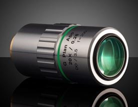
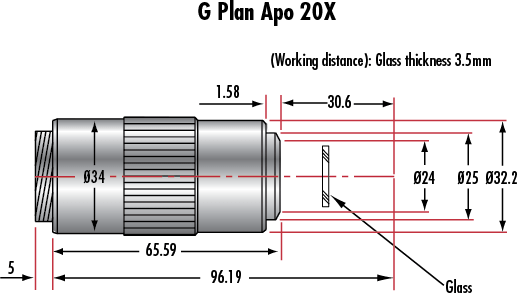
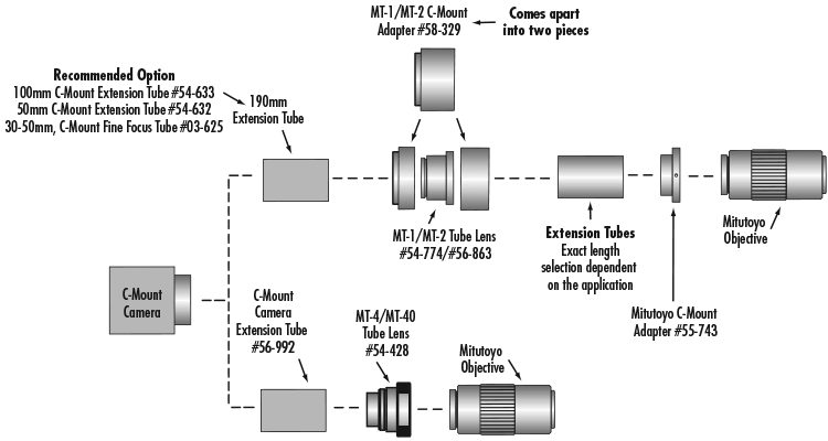
 EO Spec Sheet


もしくは 現地オフィス一覧をご覧ください
クイック見積りツール
商品コードを入力して開始しましょう
Copyright 2023, エドモンド・オプティクス・ジャパン株式会社
[東京オフィス] 〒113-0021 東京都文京区本駒込2-29-24 パシフィックスクエア千石 4F
[秋田工場] 〒012-0801 秋田県湯沢市岩崎字壇ノ上3番地