製品がカートに追加されました

SWIR Plan APO 無限補正対物レンズ 50X

SWIR Plan APO 無限補正対物レンズ

SWIR Plan APO 無限補正対物レンズ

×
商品コード #26-221 お問い合わせ
×
Quantity Selector - Use the plus and minus buttons to adjust the quantity. +
¥926,150
数量 1+
¥926,150
クイック見積り
見積依頼する

仕様

物理的および機械的特性

自重 (g):
500

光学的特性

焦点距離 FL (mm):
4.00
倍率:
50x
開口数 NA:
0.6
分解能 (μm):
1.1
被写界深度 (μm):
1.50
カバーガラス補正:
-/1.0mm(LCD)
作動距離 (mm):
10
波長範囲 (nm):
900 - 1600
最大イメージサークル (mm):
24.00

ねじ径 & 固定

固定用ねじ径:
M26

法規制対応状況

RoHS 2011/65:
適合証明書:

商品説明

  • 1600nmまでの短波赤外 (SWIR) 波長で高解像力
  • 1X ~ 50Xの倍率でラインナップ
  • 光電子放出検出, レーザーマーキング, レーザー切断に最適

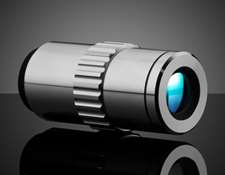
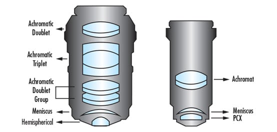
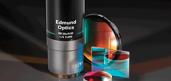
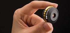
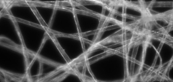
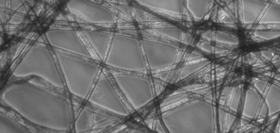
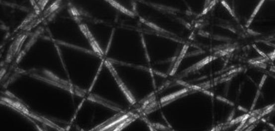
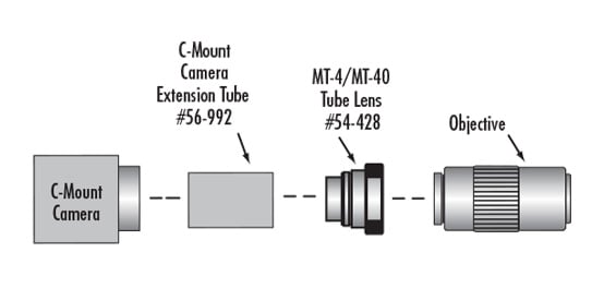
SWIR Plan APO 無限補正対物レンズは、800 – 1600nm間で色補正された長作動距離の高解像力対物レンズです。1X ~ 50Xの倍率でラインナップする本対物レンズは、SWIR波長での高解像イメージング用に高い開口数を採用します。一般的な95mmの同焦距離を持つこの対物レンズは、容易なシステム実装に向けて、焦点距離200mmの結像レンズや互換性のあるCマウントカメラと組み合わせることができます。SWIR Plan APO 無限補正対物レンズは、光電子放出検出、ウェハ裏面検査、レーザーガラス切断、およびレーザースキャン顕微鏡アプリケーションに最適です。

アクセサリー製品

タイトル 比較する商品コード  価格 (税別) カートに入れる
TS Cマウント 対物レンズ用アダプター ミツトヨ M26 #55-743 ¥6,250 数量 5+ ¥5,550   見積依頼する
  • 在庫あり
    ×
SWIR Inline Tube Lens #26-223 ¥753,500   見積依頼する
  • 5-7営業日
    ×

関連製品

一緒によく購入される商品

参考資料

Filter

顕微鏡用対物レンズの理解

Want to know more about microscopes? Learn about the different components used to build a microscope, key concepts, and specifications at Edmund Optics.

今すぐ見る

Multiphoton Microscopy

Multiphoton microscopy is ideal for capturing high-resolution 3D images with reduced photobleaching and phototoxicity compared to confocal microscopy.

今すぐ見る

Confocal Microscopy

Confocal microscopy provides high resolution, elimination of out-of-focus glare due to spatial filtering, and reduction of light-induced damage to the sample.

今すぐ見る

Infinite Conjugate Tube Length

無限補正対物レンズの分解能と倍率の理解

Microscopes are used in a variety of fields and applications. To understand how resolving power, magnification, and other aspects work, read more at Edmund Optics.

今すぐ見る

結像レンズを無限補正対物レンズと使用する

Want to use an infinity corrected objective to make an image? You will need a tube lens to do it! Find out why and how it works at Edmund Optics.

今すぐ見る

What is an "in-line" microscope?

What is an infinity-corrected objective?

Can I use an infinity-corrected objective in my microscope to get a longer working distance?

What is "DIN" and "JIS"? What is the difference between them?

I’m using the MT-1 Accessory Tube Lens and MT-1/MT-2 C-Mount Adapter. Two thin brass rings came with the adapter. How are they used?

What is the significance of the field number of an objective and what is it dependent on?

Plan Objective

Semi-Plan Objective

Tube Lens

顕微鏡用対物レンズの未来

コンパクトで小型化された対物レンズは、顕微鏡システムをポータブルにすることができ、現場での迅速な作業を可能にします。

今すぐ見る

Optical Microscopy Application: Brightfield Illumination

Looking to use a simple technique for optical microscopy? Check out this article on brightfield illumination to find out more at Edmund Optics.

今すぐ見る

Optical Microscopy Application: Darkfield Illumination

Darkfield illumination is the opposite of brightfield illumination. Find out how darkfield differs from brightfield in optical microscopy at Edmund Optics.

今すぐ見る

Optical Microscopy Application: Phase Contrast

Think you know all the advantages for using phase contrast in optical microscopy? Advantages, image appearance, and technical details can be found at Edmund Optics.

今すぐ見る

What is the difference between Achromatic, Semi-Plan and Plan objectives?

You have a variety of Mitutoyo Tube Lenses (MT-4, MT-1, and MT-2). How can you connect them to a Mitutoyo objective and C-mount camera?

I have an MT-L Accessory Tube Lens but I am unsure about mounting it to my objective and C-Mount camera. Can you explain how it works?

Achromatic Objective

Apochromatic

Confocal Microscopy

DIN (Deutsche Industrie Norm)

Periplan Eyepiece

光学ケージシステムアプリケーション: デジタルビデオ顕微鏡

Are you looking to simplify the assembly of a digital video microscope? Find out how a TECHSPEC Optical Cage System allows for quick build at Edmund Optics.

今すぐ見る

Optical Microscopy Application: Differential Interference Contrast

Differential interference contrast (DIC) is one of the polarization techniques that can be used in optical microscopy. Learn about this technique at Edmund Optics.

今すぐ見る

Field Number

Light Sheet Microscopy

Light sheet fluorescence microscopy uses a 2D laser sheet to illuminate a thin slice of the sample and excite fluorescence, reducing phototoxicity and damage.

今すぐ見る

Optical Cage System Application: Differential Interference Contrast (DIC) Digital Microscope

Differential interference microscopy enhances the contrast of object features that are otherwise difficult to observe using brightfield microscopy.

今すぐ見る

Darkfield Illumination

Diffraction Limit

Parfocality

デジタルビデオマイクロスコープのシステムセットアップ

Digital video microscopes use a camera to capture and record images. Read more about the components needed to assemble a video microscope at Edmund Optics.

今すぐ見る

Optical Microscopy Application: Fluorescence

Wondering how fluorescence microscopy works? Find out about the technique, systems, and more at Edmund Optics.

今すぐ見る

Brightfield Illumination

Field of View (FOV)

Magnification

Numerical Aperture (NA)

蛍光顕微鏡:画像用フィルターを用いた同軸落射照明

Want to know about fluorescence microscopy? Read this article by a Biomedical Product Line Engineer at Edmund Optics to learn more.

今すぐ見る

Refraction

 
販売や技術サポート
1-800-363-1992
もしくは 現地オフィス一覧をご覧ください
簡単便利な
クイック見積りツール
商品コードを入力して開始しましょう
This site is protected by VikingCloud's Trusted Commerce program